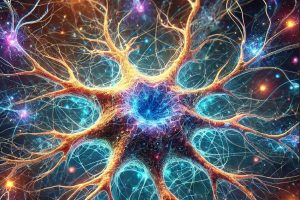
Astrocytes—the star-shaped cells that provide nutrients to nerve tissue, regulate neurotransmission, and aid in repairing brain and spinal cord scarring—are crucial yet understudied. An international research team led by Ishan Barman, a professor of mechanical engineering at the Whiting School of Engineering, has leveraged a new imaging system to better explore astrocyte behavior, marking the first use of this method to visualize this specific type of neural cell and opening new avenues in neuroscience research. The research appears in Advanced Healthcare Materials.
Called Optical Diffraction Tomography (ODT), the new system captures the changes in the shape and structures of astrocytes over time— an aspect that has been underexplored—without using any added dyes or chemical markers to make cells visible.
ODT reconstructs three-dimensional maps showing how light bends through cells, providing crucial insights into their structure and composition. By capturing multiple images of cells from different angles and computationally reconstructing a 3D image, ODT allows researchers to visualize subtle changes in cellular morphology with high precision.
“This study demonstrates the power of ODT to reveal intricate morphological changes in astrocytes without disrupting their natural behavior,” Barman said. “By capturing real-time, quantitative data, we are now able to track how these cells evolve and interact within neural networks.”

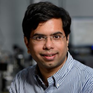
Ishan Barman
The team’s system improves on previous imaging methods by providing real-time, high-resolution insights into cell volume, dry mass, and surface area without the need for fluorescent molecules, which can be detected by an optical microscope but may also alter cell behavior. The study also introduces a novel computational approach for tracking astrocyte development, which allows researchers to map their transitions, offering a new framework for analyzing neural cell interactions.
“Understanding how astrocytes change their shape and structure over time gives us a new lens into their function in both healthy and diseased states,” said Pooja Anantha, the study’s first author and a graduate student in the Barman laboratory.
Other Hopkins contributors include Jeong Hee Kim and Piyush Raj, from mechanical engineering, and Assistant Professor Luo Gu and Joo Ho Kim, from materials science and engineering. Emanuela Saracino from CNR-ISOF in Bologna and Annalisa Convertino from CNR-IMM in Rome were also team members.
The research was supported by the Air Force Office of Scientific Research (AFOSR) Biophysics Program