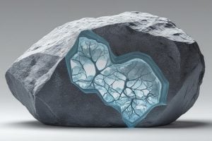
It seems like a fundamental concept: squeeze a rock and its internal structure changes until it ultimately breaks. But geologists have never been able to see in three dimensions exactly how individual grains move and stress builds up inside that rock— until now. Using an innovative X-ray imaging approach, a team of Johns Hopkins researchers has revealed how compression reshapes the tiny spaces and stresses within sandstone—findings that could predict how this common rock used for fuel reservoirs behaves under deep subterranean pressure.
“We want to understand how forces are transmitted through rocks and how that transmission changes as you increase the force and eventually break the rock,” said Ryan Hurley, associate professor of mechanical engineering and a research fellow at the Hopkins Extreme Materials Institute. “Why? Because these processes govern everything that happens in the Earth’s crust, from our man-made activities like oil reservoir stimulation to natural events like earthquakes.”
The study, one of the first of its kind, was published in the Journal of Geophysical Research: Solid Earth and was an Editor’s Highlight in the science news magazine Eos.
In their investigation, Hurley’s team used tools, some of which are used for medical imaging, to peer inside Nugget sandstone, generally found in the American West, examining its complex network of pores, grains, and pockets, which dictate how a break occurs.

Ryan Hurley
The researchers used a suite of X-ray measurement techniques: X-ray tomography, which provides 3D images of a rock’s structure; 3D X-ray diffraction, which can see stresses in each grain throughout a rock; and near-field high-energy diffraction microscopy, which shows the orientations of the crystals of each grain within a rock. These methods have been used by materials scientists to study metals, but Hurley said his group is among the very few—and firs—to use them all in tandem and specifically to study rocks.
“Initially, we used these techniques to study simple materials composed of collections of single crystals, but now we’re using all of the techniques together to build a complete picture of a rock’s structure, crystalline texture, and force transmission during mechanical loading,” he said.
Hurley explained that the largest determining factors that dictate how much stress a rock can take before breaking are texture—the crystal orientation of grains—and structure—where the grains and voids are. The researchers saw what they called the “stress evolution” within the rock, observing that behavioral links existed between rocks and non-cohesive granular materials like sand and gravel, with both having similar reactions to external stresses. Additionally, as the sandstone was compressed, its pores closed in the direction of the force and opened up sideways, which caused the sample to crack but not fully break apart.
“These studies reveal that rocks under stress may, in certain circumstances, behave like collections of interacting grains, exhibiting similarities with inter-particle force transmission in granular materials,” Hurley said. “This suggests a link that has been proposed for over a decade but not, until now, confirmed.”
Hurley said this work provides a proof-of-concept for using the same suite of techniques to study the relationship between texture, structure, and mechanics of rocks in exquisite detail.
“With advances in-situ X-ray capabilities emerging in the next few years, we intend to use these techniques to study larger samples in application-relevant stress conditions, like those present in subsurface reservoirs or near faults,” he said. “We also plan to develop and validate ‘digital twins’ of our samples and study how alterations in structure or texture change mechanics, so that our findings can apply as broadly as possible.”